Stemcelldoc's Weblog

November 20, 2020

Do You Need Surgery for Craniocervical Instability

Filed under: Uncategorized — stemcelldoc @ 2:54 pm


https://centeno-schultz.buzzsprout.com/817513/6404431-do-you-need-surgery-for-craniocervical-instability

At Centeno-Schultz Clinic, we host a smaller operation called CCJ Instability Institute where we are one of the very few who successfully treat craniocervical instability without surgery. https://centeno-schultz.buzzsprout.com/817513/6404431-do-you-need-surgery-for-craniocervical-instability

Please enjoy the podcast. If you have questions, please contact Centeno-Schultz Clinic @ https://centenoschultz.com/contact/

April 29, 2014

Successful Treatment of AVN of Femoral Head with Stem Cell Therapy

Avascular necrosis of the femoral head is bone death thought to arise from interruption of the blood supply.

Progression of the disease is characterized by flattening of the femoral head with eventual collapse of the hip joint.

Stem cell therapy is a non surgical option in the treatment of AVN of the femoral head.

MFH is a 50y/o active female initially evaluated at the Centeno-Schultz Clinic with ARCO stage 2 AVN of the femoral head.

On 4.2009 she underwent core decompression where bone marrow derived stem cells were injected directly into the area of necrosis.

Clinically patient has done extremely well as reflected in her 5 year follow up questionnaire which she permitted us to share.

Her hip x-rays demonstrate successful treatment of AVN with Regenexx treatment.  Note that the contour of the femoral head has not changed, flattened or collapsed.

MFH AVN

April 27, 2014

Successful Platelet Epidural Treatment for Severe Leg Pain

Helicopter skiing in Canada.

helicopter skiing canada

Skiing the trees in Aspen.

Surfing in California.

california-surfer


These are activities that BC enjoyed after undergoing specialized platelet epidural injection at the Centeno-Schultz Clinic.

Regenexx PL-Disc utilizes a supercharged collection of platelet growth factors that are injected under x-ray around the irritated nerve roots and lumbar disc.

Regenexx PL-Disc is differs from traditional platelet rich plasma (PRP) as it created by a cell biologist, has reduced white and red cell contamination and higher concentration of platelet growth factors.

High steroid epidural steroid injections have significant side-effects and platelet injections are a viable alternative as discussed in video below.


CB is  39 y/o outdoor enthusiast who presented to the Centeno-Schultz Clinic with a 20 year history of right leg pain that was progressive in nature originating in the buttock extending down to the lateral aspect of the foot.   Aggravating factors included coughing, sneezing and prolonged sitting.   Treatment to date had been extensive and included heat, ice, physical therapy, massage, chiropractic care and high dose epidural steroid injections.  Patient had undergone a L4/5 laminectomy and decompression due to disc herniation in 1991.

MRI was significant for facet hypertrophy and right-sided disc bulge at L4/5 with impingement of the L4 nerve root.

CB underwent two x-ray guided injections of the right L3-S1 facets and the right L4 and L5 nerve roots and epidural space with Regenexx PL-M over 2 months.

These treatments allowed him to engage in a 14 day 3 state epic trip.

I saw CB in clinic post trip and he continued to feel great with no leg pain or restrictions.

Congratulations !


April 21, 2014

ITB: Iliotibial Band Dysfunction and Treatment Options

At the Centeno-Schultz Clinic we approach pain and dysfunction in a systematic fashion which is outlined in Ortho 2.0.

We employ the acronym SANS:  symmetry, articulation, neurologic and stability.

This approach can be utilized for any joint.

The iliotibial band (ITB) is a fibrous band that extends from the hip (ilium) to the knee (tibia).

It is a critical in the stability of the hip and pelvis.

There are both anterior and posterior forces on the ITB.

Posteriorly it connects with the gluteus medius  muscle which when contracted pulls the ITB posteriorly.

Anteriorly the ITB connects with the tensor fasciae latae muscle which when contracted pulls the ITB anteriorly.

TFL_ITBand

Biomechanics of ITB:  Supports and stabilizes the hip and pelvis when the opposite foot is suspended and off the floor such as when we are walking.

Lumbar spinal nerves provide essential information to the gluteus medius, tensor fasciae latae and other muscles that support the ITB.

Irritation or compression of lumbar nerves can result in a reduction in the nerve signal to the muscles and ensuing weakness.

This weakness can be witnessed on physical examination when the patient is asked to stand on one leg and the pelvis shifts.  Weakness in the ITB compromises the stability of the pelvis and hip with resultant shift of the pelvis as illustrated below.


ITB weakness


An excellent analysis and summary of the iliotibial band is presented below by Dr. Centeno.


April 17, 2014

Anterior and Posterior Cruciate Ligament Injection of Stem Cells Under X-Ray

At the Centeno-Schultz Clinic accuracy is a cornerstone of our practice.  Accordingly we perform all injections under either x-ray or MSK ultrasound guidance.

The Anterior and Posterior Cruciate ligaments (ACL and PCL) are critical stabilizers of the knee.

LL is a 35 y/o athletic male from Indonesia who sustained tears into both the ACL and PCL and rejected traditional surgery.   Today we injected bone marrow derived stem cells into both ligaments with precision utilizing x-ray guidance.  Below is the image demonstrating the injection of contrast into both the ACL and PCL.  a small amount of contrast is injected prior to the stem cells to confirm accurate needle placement.  The PCL is outlined in yellow whereas the ACL is in red.

Anterior and Posterior Cruciate Injection


April 10, 2014

Avascular Necrosis of Femoral Head: Stem Cell Treatment Options

Avascular necrosis (AVN) is bone death thought to arise from interruption of the blood supply.  The MRI below illustrates bone death (AVN) in the left hip characterized by irregular shape of the femoral head and dark black bone.  This is in contrast to the normal hip on the right.

Avascular-necrosis-(AVN)2


Stem cell therapy is a non surgical option in the treatment of AVN of femoral head.

Core decompression is a x-ray guided procedure that places bone marrow derived stem cells directly into the area of necrosis.

Hernigou demonstrated the clinical efficacy of core decompression with bone marrow derived cells in the treatment of hip avascular necrosis.  534 patients with early stages of AVN were treated and best clinical outccomes were noted in patients without collapse of the femoral head (stage 3).

 Prior blog posts have discussed clincial successes.

Common causes of AVN include excessive alcohol, steroid use, trauma, vascular compression and chemotherapy.

Classical presentation involves the head of the femur, neck and talus and scaphoid.

Typically affects individuals between 30 and 50 years of age.

The two most commonly used classification system used are the Ficat andArlet Staging and ARCO staging.

AVN staging

A cardinal finding is the crescent sign which is seen on x-ray and refers to a linear area of subchondral lucency most frequently in the anterolateral aspect of the proximal femoral head.  The sign indicates a high likelihood of collapse.  The crescent sign is best seen in frog leg position (abduction).

crescent sign

Prognosis depends upon severity of the bone death.

Non surgical options utilizing bone marrow derived stem cells are available for patients with avascular necrosis at the Centeno-Schultz Clinic.

April 6, 2014

Treatment of Knee Arthritis Pain: Curcumin vs NSAID

At the Centeno-Schultz Clinic we understand the limitations, physical and emotional drain associated with pain.

Curcumin is an active ingredient in the Regenexx Advanced Stem Cell Support Formula.

It is an innovative formula of nutritional supplements that helps maintain and support normal joint health and functioning.  In Regenexx laboratory studies, the supplements within the formula helped to maintain the health of the cell environment and provide support for the joint’s natural cartilage growth (chondrogenesis) process.

What is Curcumin?

tumeric_0706

Curcumin an extract from the Indian spice tumeric.  Research has shown curcumin to modulate inflammation.

Is Curcumin as effective a Ibuprofen?

Yes!

Kuptniratsaikul et al recently compared the efficacy and safety of Curcumin with Ibuprofen in patients with knee osteoarthritis.  In a multicenter study, 367 patients with knee osteoarthritis were randomized to receive 1,200 mg/day of ibuprofen or Curcuman extracts 1500mg/day for 4 weeks.  Curcumin was as effective as Ibuprofen in improving pain and function in patients with knee osteoarthritis.  While the number of adverse events was not different between the groups, there was a higher number of abdominal pain/discomfort in the ibuprofen group.

Are there other differences?

Absolutely!!

The chart below outlines the key differences.

NSAID have significant side effects which include reduced bone healing, increases in systematic inflammatory markers, increases in oxidative stress and increases in deadly heart attack risk by 200-300%.

Know the risks and natural alternatives for management and treatment of knee arthritis pain.

knee-arthritis-supplement

April 5, 2014

Saphenous Nerve Entrapment as a source of knee pain

At the Centeno-Schultz Clinic we acknowledge that there can be many causes of knee pain.

In our online book, Ortho 2.0 a systematic approach is discussed: SANS.

N references neurologic dysfunction.

Irritation or dysfunction of the saphenous nerve can be cause of knee pain.

The saphenous nerve is a pure sensory nerve compromised of fibers from L3 and L4.  The saphenous nerve is the longest branch of the femoral nerve.

femoral nerve division

Branches

Above the knee joint the infrapatellar branch arises whereas below the knee the medial crural cutaneous branch provide sensation to the front and medial aspect of the knee.

Dermatomes of Saphenous Nerve

A common site of entrapment is adductor (Hunter’s) canal which is an aponeurotic tunnel in the middle third of the thigh.  Adductor canal contains femoral artery, femoral vein and branches of the femoral nerve which include the saphenous nerve.

The Adductor canal has the following boundaries:

  • Anteriorly – Sartorius
  • Postermedially – adductor longus and adductor magnus
  • Laterally -vastus medialis

adductor canal

adductor canal

Ultrasound image of saphenous nerve in Adductor canal

saphenous nerve ultrasound image adductor canal

A systematic approach with diagnostic tools that include MSK ultrasound ensure maximal patient outcomes.  Treatment options of saphenous nerve irritation/entrapment include MSK ultrasound guided hydrodissection with platelet growth factors.

March 30, 2014

Lateral Arm Numbness and Weakness: Consider Axillary Nerve Dysfunction

At the Centeno-Schultz Clinic we acknowledge that shoulder pain can compromise quality of life and athletic endeavors.

Our online book, Ortho 2.0 highlights a systemic evaluation of joint pain and dysfunction.  The approach is termed SANS.

The N is SANS denotes nerve dysfunction.

Axillary nerve dysfunction can be a cause of lateral arm numbness and shoulder weakness.

The Axillary nerve arises from the posterior cord of the brachial plexus, C5 and C6.

Course:  It crosses the antero-inferior aspect of the subscapularis muscle, passes behind the arm through the quadrilateral space, winding around the neck of the humerus ending in two major trunks:  anterior and posterior trunk.

Axillary nerve

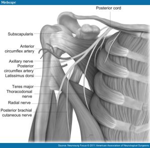
Innervation:  Teres Minor and Deltoid

Sensory: Lateral deltoid also known as the regimental badge area.

Axillary-Nerve

Injury can occur at several sites along the nerve path:

Origin of the posterior cord

Anterior-inferior aspect of the subscapularis muscle

Quadrilateral Space:

suprascapular_and_axillary_nerves_02

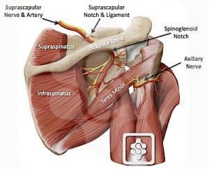
The quadrilateral space is bounded superiorly by the teres minor muscle, inferiorly by the teres major muscle, medially by the long head of the triceps, and laterally by the humeral shaft.

Common presentation is dull intermittent ache or pain that is localized in the posterior and lateral shoulder.  Symptoms are exacerbated by active and resisted abduction and external rotation of the humerus.

Quadrilateral space syndrome is an affliction of middle-aged men and it presents classically in the throwing athlete and those with a history rotator cuff surgery.

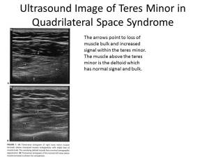
Ultrasound examination is can be significant for atrophy of the teres minor muscle which is illustrated below.

Ultrasound Image of Teres Minor in Quadrilateral Space

 injury at several sitesgin from the posterior cordAnteroinferior aspect of the subscapularis muscle and shoulder capsuleQuadrilateral space

March 26, 2014

Common Causes of Chronic Knee Pain: A systematic Approach

At the Centen0-Schultz Clinic we utilize a comprehensive approach to patient care.    Our online ORTHO 2.0 discussess a systematic approach to common orthopedic conditions.

Don’t assume that loss of cartilage is the source of all knee pain.

Below is an infographic which outlines the common causes of knee pain.

Tendons:  patellar, distal quadriceps and pes anserine.

Spine:  Lumbar disc disease with compression of L5 and or S1 nerve root.

Peripheral nerves:  compression and or irritation of saphenous and tibial nerves.

Muscles:  Dysfunctional muscles resulting in weakness or imbalance.

Ligaments:  key stabilizers of the knee joint include the ACL, MCL and LCL.

Knee Joint:   medial and lateral meniscus

Bottom Line:  prior to undergoing any treatment ensure that the principal pain generator has been identified to maximize clinical results.


Common-causes-of-Chronic-knee-pain-Infographic

Older Posts »